3D MAMMOGRAPHY OVERVIEW

What is breast tomosynthesis?

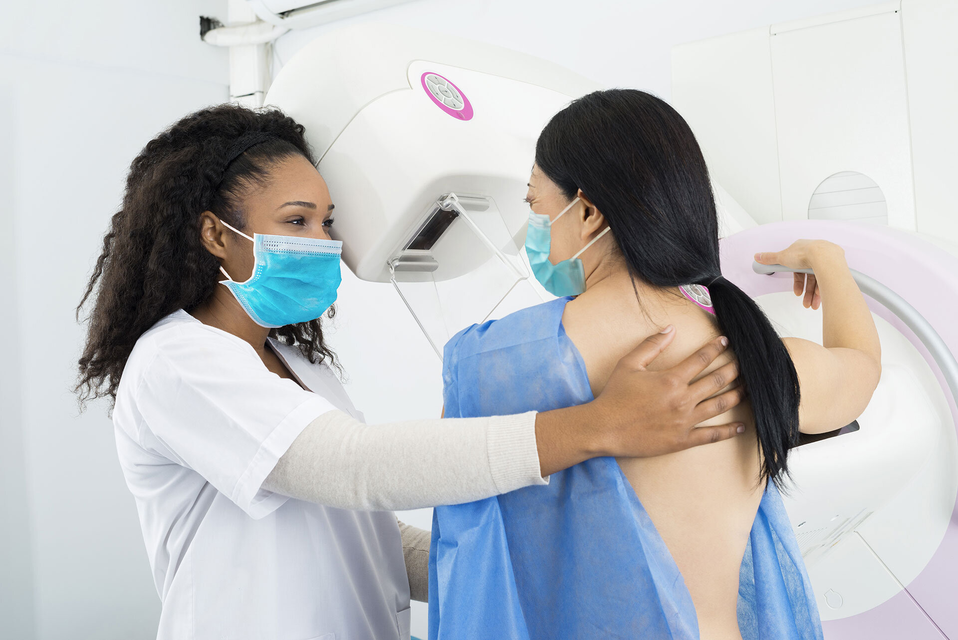
Mammography remains the “Gold Standard” for breast cancer screening and early detection. Breast tomosynthesis, or 3D mammography, represents a significant breakthrough in breast imaging technology, allowing your radiologist to view your mammogram at a level detail that simply wasn’t available in the past.

Tomosynthesis produces a three-dimensional picture of the breast that a radiologist can view in 1-millimeter slices. The additional 3D images allow radiologists to provide a more comprehensive evaluation of a patient’s breast tissue during screening while reducing the need for follow-up imaging.

In studies where the 2D and 3D images are both obtained, the total X-ray dosage is still below the safety limit by the Mammography Quality Standards Act (MQSA) set forth by the FDA.

What are the advantages of 3D mammography?

A significant research study reported in the JOURNAL OF THE AMERICAN MEDICAL ASSOCIATION found that tomosynthesis imaging, or 3D mammography, reveals significantly more invasive cancers than a traditional, 2D mammogram. Invasive cancers are more likely to spread or cause death. 3D mammography also reduces the number of women called back for additional imaging, which reduces patient anxiety and helps lower health care costs.

The study’s findings included:

  • 41% increase in the detection of invasive breast cancers

  • 29% increase in the detection of all breast cancers

  • 40% decrease in false positives

  • 15% decrease in women recalled for additional imaging

How do I get a 3D mammogram? To schedule your annual screening mammogram, call Inland Imaging at 509.455.4455.

BREAST ULTRASOUND and automated breast ultrasound (abus)

Ultrasound uses sonar, or high-frequency sound waves, to capture real-time images of organs inside the body. The noninvasive and generally painless exam is often used to determine if a suspicious area is a fluid-filled cyst or a solid mass that requires further testing. The imaging tool is also used to guide breast biopsies.

What’s an Automated Breast Ultrasound?

Approximately 40% of women have dense breasts, one of the strongest common risk factors for developing breast cancer.  Having dense breasts may increase a woman's chance of developing breast cancer. A high percentage of breast cancers are found in women with dense breasts.

2D and 3D mammography may miss some cancers in dense breasts, potentially delaying diagnosis. Both dense breast tissue and cancer appear white on a mammogram, sometimes creating a dangerous camouflage effect and a dilemma for radiologists whose goal is to find breast cancer as early as possible.

Automated Breast Ultrasound is a comfortable alternative to other supplemental screening options for women with dense breast tissue. When used in addition to mammography, ABUS was shown to improve breast cancer detection by 37.5 percent over mammography alone in a recent study. 

What to expect?

For most breast ultrasound exams, you will lie face-up or on your side. A clear gel is applied to the skin to eliminate air pockets between the transducer and the skin. An ultrasound technologist firmly presses the transducer against your skin, moving it back and forth over the specific area.

Generally the exam is completed within 15-30 minutes. In some cases, at the conclusion of your exam you may be asked to dress and wait while the ultrasound images are reviewed. Breast ultrasound is used frequently in conjunction with diagnostic mammography to help identify and clarify clinical and mammographic findings.


BReaST MRI and abbreviated breast mri

Unlike mammography, which uses low-dose X-rays to image the breast, MRI uses powerful magnetic fields and radio waves to create images of the breast.

Biopsies may also be performed using breast MRI. MRI-guided breast biopsy is a fast, safe and easy way to find and biopsy breast abnormalities without putting women through unnecessary surgery. Our state-of-the-art breast MRI system optimizes patient comfort, reduces unnecessary movement during the exam, and produces superior images. It also offers improved access for biopsies.

What’s an Abbreviate Breast MRI?

“AB-MRI,” also called abbreviated MRI or fast MRI, is a shortened version of breast MRI that is designed to screen for additional breast cancers not seen on mammography.

AB-MRI detects biologically aggressive invasive cancers at early stages. The sensitivity of AB-MRI is comparable to that of conventional MRI.

What should I expect?

For a breast MRI exam, you will lie face down on the specially designed breast MRI table, which is configured to allow the breasts to be positioned comfortably through two openings called breast coils. Sometimes, patients are given a contrast material through IV, which improves viewing of the targeted area.

A Breast MRI without IV Contrast takes approximately 45 minutes. For a Breast MRI with IV contrast, expect the exam to take 45-50 minutes.


BREAST BIOPSY

If your mammogram showed an abnormality in your breast, a breast biopsy may be your next step. This non-surgical procedure involves using a biopsy needle to remove a small sample of tissue from the area of concern. The results will help your physician determine if that suspicious-looking spot on the mammogram is going to need more attention.

Imaging-guided breast biopsy is often an excellent alternative to a surgical biopsy. Performed on an outpatient basis, the procedure is accurate, cost-effective (one-third the cost of surgery), does not require hospitalization or general anesthesia, and does not cause significant scarring.

What to expect?

With the guidance of breast-imaging technology, a specially trained radiologist will insert a thin, hollow breast-biopsy needle into the area of concern to remove a small sample of the suspicious tissue. A local anesthetic is applied to numb the area.

Depending on your mammogram results, the biopsy may be guided by stereotactic (X-ray) technology, ultrasound or MRI. A typical biopsy takes approximately one hour.

Following the biopsy, you should avoid strenuous exercise, heavy lifting, reaching, pulling or tugging for 48 hours. You will receive additional post-care instructions after the procedure.

Receiving your results.

Your biopsy results will generally be available from the pathology laboratory within 72 hours. Be sure to review the results with your physician.


BREAST WIRE AND WIRELESS LOCALIZATION

Localization procedures allow our radiologists and technologists to accurately locate an abnormality that will later be surgically removed. Using the appropriate imaging modality, a localization wire is placed in the breast to accurately target suspicious tissue that will be surgically removed later that same day.

In other cases patients may be able to use wireless location technology that uses radar to target the anomaly, which has been marked by the insertion of a tiny reflector. This can be done at the patient’s convenience days before their surgery is scheduled.

During surgery, surgeons can electronically access the images showing the location of the targeted cells. Imaging tests are done on the surgically excised specimen to verify removal of the abnormality. Results are then phoned to the surgeon.