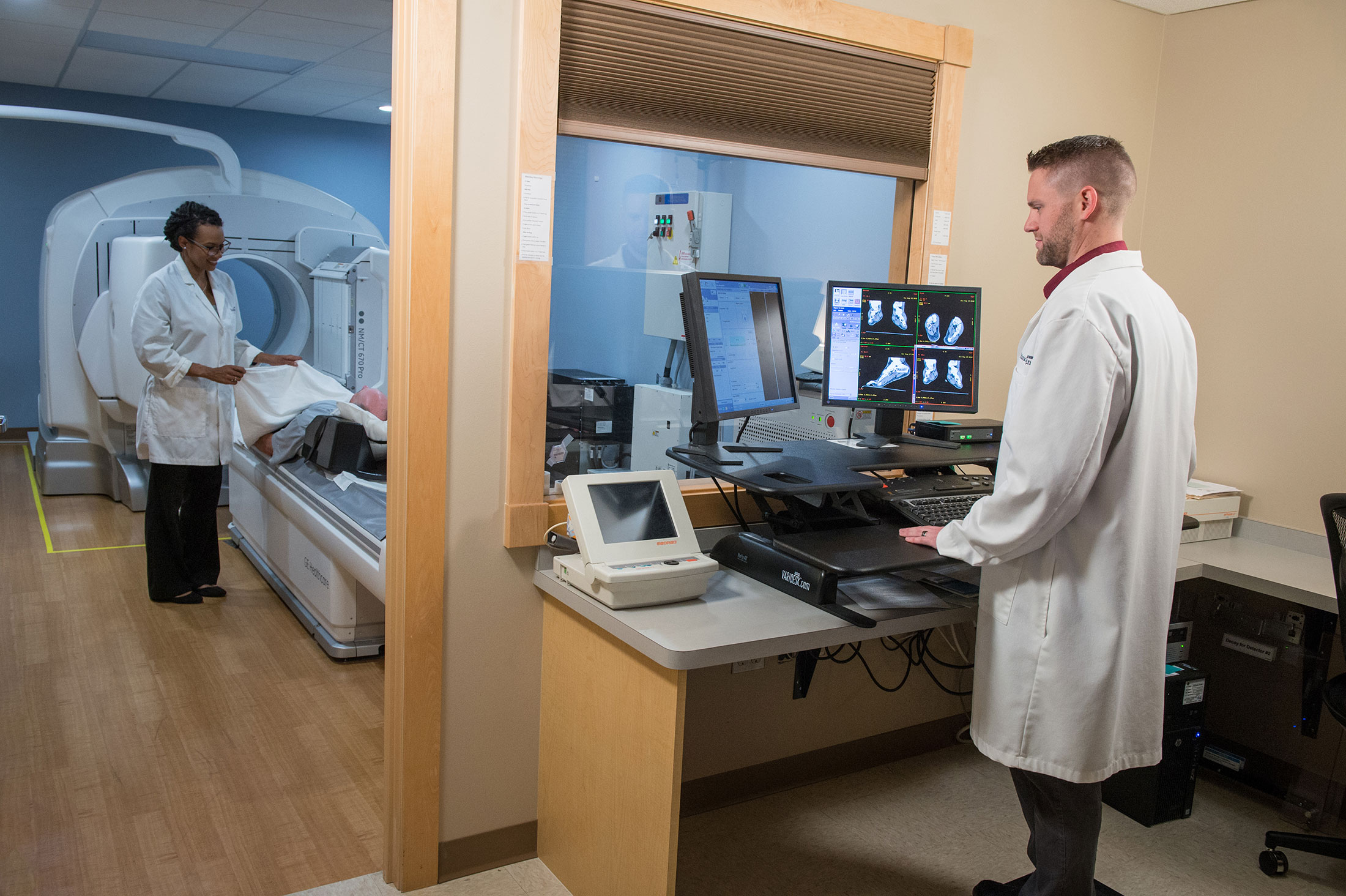
BONE IMAGING

A bone scan is a nuclear imaging exam that can help physicians diagnose and treat problems of the bone, such as cancer, arthritis, fractures, infections and other conditions. This test uses a small amount of a radioactive fluid, called a radioisotope, which is absorbed by the bones over time. The gamma camera detects energy from the radioisotope and produces functional images of the skeleton. 

What should I expect?

When you first arrive for your bone scan, the technologist will inject a small amount of radioactive material into your vein.

You must drink at least 2 to 4 glasses of water or juice within the first hour following your injection.

After your injection, it takes 2 ½ to 4 hours for the radioactive material to travel to your bones. You can leave the facility during this time. You will be given a time to return for your scan which will take approximately 45 minutes” or something similar.

BRAIN SCAN

An imaging exam using nuclear medicine can help your physician see and evaluate brain blood flow and function. This information is helpful in the diagnosis of conditions such as dementia and other neurological conditions of the brain.

What should I expect?

An IV line will be placed in your hand or arm to administer the radioactive scanning drug. Depending on the reason for the scan, you may rest quietly in a low-lit room for 45 minutes before your scan. Sometimes there will be a three-hour delay from the injection time to the scan time. You may leave during these three hours and return for your scan at the time givento you by your technologist.

During the scan, the technologist will position a gamma camera near your head to take pictures of your brain. The imagingtakes approximately 30 minutes.

CISTERNOGRAM

A cisternogram is a nuclear imaging test that shows the distribution of the cerebrospinal fluid, also known as CSF

What should I expect?

Your back will first be numbed locally with an anesthetic. You may feel some pain as the area is numbed. Next, the radioactive material will be injected into your spinal column with a needle. You may feel slight pressure as the needle is inserted into the lower back. The needle will be removed after the injection.

Scans of your back and head will be taken at various intervals of time. Typically, imaging sessions will take place at three hours, 24 hours, 48 hours, and, sometimes, 72 hours after your injection.

GALLIUM SCAN

A gallium scan is a nuclear imaging test that uses gallium, a radioactive substance that is designed to build up at sites of tumors or areas of chronic infection. A scan will show these problem areas as bright spots on an image and provide information that may help your physician detect, diagnose and treat cancer, infection or inflammation inside the body.

What should I expect?

A gallium scan often requires multiple visits to our facility for imaging. During your first visit, your technologist will inject a small amount of gallium into your vein. After your injection, you will be asked to return one, two or three days later for your imaging test. You will be asked to return for imaging at 24 and 48 hours after the injection and rarely you may be asked to return for a 72 hour scan.

GASTRIC EMPTYING

A gastric emptying test is performed to determine the time it takes for solid food to empty from your stomach into your small bowel.

What should I expect?

The standard meal for the gastric emptying exam consists of eating two scrambled eggs, two pieces of white bread with strawberry jam, and 5oz of water.  Once you are done eating the technologist will take a two-minute picture every thirty minutes for two hours.  After the two-hour image is done you may leave the facility for just under two hours but you are not allowed to eat or drink anything during that time.  You must return for one final image four hours after the first image was taken.  If you are not back promptly at the four-hour mark you may have to repeat the exam at your own expense due to lack of accuracy of the results.  Sugar-free jam is available upon request.  If you are gluten intolerant, please bring two pieces of gluten-free bread with you to the appointment.  If you have an egg allergy or if the amount of food listed in the standard meal is too much for you to consume we will use 8oz of Boost Plus which is a meal replacement shake.  If your doctor orders a gastric emptying test with Reglan, once you are done eating the meal we will take pictures constantly for 90 minutes then give the Reglan and take pictures for another 30 minutes.

How should I prepare?

Nothing to eat or drink 12 hours prior to the exam. May have water up to 4 hours prior to appointment. Do not take narcotic pain medications for 24 hours prior to the exam. Do not take narcotic pain medications for 24 hours prior to the exam. If you are currently taking narcotic pain medications please tell the scheduler.

GASTROINTESTINAL BLEED

A nuclear imaging scan of your gastrointestinal (GI) system can help your physician find the source of bleeding in your intestines.

What should I expect?

This drawn blood is mixed with a radioisotope in the lab that binds to your red blood cells.  This process is called labeling. The treated blood is injected back into the vein in your arm and allows us to image where the red blood cells go in your body.

Once the radioactive blood is in your vein, the technologist will take pictures of your abdomen over a period of approximately 60 minutes.

Your appointment is usually scheduled for the morning. Your initial visit will take approximately two hours. Sometimes, you will be asked to return later in the day for additional pictures, which may require an additional 30 minutes.

HEPATOBILIARY SCAN

A hepatobiliary scan is a nuclear imaging exam that will show if there are blockages in your gallbladder or bile ducts. If your gallbladder has been removed, the test may be performed to detect leaks in the bile ducts and delayed bile flow into the small bowel.

What should I expect?

Expect to spend an average of 2.25 hours for this appointment.

A small amount of radioactive material will be injected into your body. This radioactive material will show the flow of biliary fluid. A technologist will begin taking pictures immediately after your injection. This initial image session may last up to an hour. If the technologist is unable to view the flow of the biliary fluid during that time, imaging will continue intermittently for up to an additional 3 hours.

Once the gallbladder is full of the radioactive tracer the technologist will give you a slow infusion of a hormone called CCK to make the gallbladder contract.  If CCK is not available they may also use 8oz of Boost Plus.  Calculating how much the gallbladder contracts after being stimulated is what tells us if the gallbladder is working properly or not.

How should I prepare?

Do not eat or drink anything four hours prior to your exam.  Within 24 hours of your exam, you must eat a fatty meal.  This could be a hamburger, fried food, ice cream, salad with extra salad dressing on it, etc. The fatty meals cause the gallbladder to contract that evening which usually helps it absorb the medicine we give you for the exam faster.  If you are unable to eat a fatty meal please tell the scheduler when you make the appointment as extra time may need to be added to your exam.

Do not take narcotic pain medications for 24 hours prior to the exam. If you are currently taking narcotic pain medications please tell the scheduler.

I-123 MIBG

An I-123 MIBG scan is a nuclear imaging exam that helps physicians find adrenal gland tumors in the body.

What should I expect?

A radioactive material will be injected into your body. Pictures are taken at 24 hours and possibly 48 hours following your injection. Prior to the exam and sometimes after your exam, you will be given drops that block the thyroid from absorbing the radioactive material.

How should I prepare?

You may be asked to stop taking certain medications prior to the study.

LIVER AND SPLEEN SCANS

A nuclear imaging exam can help your physician see the size of your liver and spleen and evaluate how well these organs are functioning. This information can help in the diagnosis and treatment of injury or disease.

The test may also be performed to detect a hemangioma, a benign tumor generally found in the liver.

What should I expect?

Your technologist will inject a small amount of radioactive material in a vein. After your injection, a gamma camera will be use to take images of your body from many different angles. The scan typically takes about 45 minutes.

For an exam looking for a hemangioma in the liver, your technologist will first draw a small amount of blood from a vein in your arm. This drawn blood is mixed with a radioisotope in the lab that binds to your red blood cells.  This process is called labeling. The treated blood is injected back into the vein in your arm and allows us to image where the red blood cells go in your body.

LUNG SCANS

A nuclear imaging scan of your chest will show how well air and blood is flowing through your lungs.

What should I expect?

A lung ventilation test will be performed to test the airflow through your lungs. In this procedure, you will breathe in a small amount of radioactive aerosol for five minutes. Images will be taken of your chest from various angles during this time.

A second set of pictures will be taken after a small amount of radioactive material is injected into an arm vein. These pictures will be taken at multiple angles to evaluate areas in the lung where blood supply is weak.

This exam takes approximately 60 minutes.

MUGA SCAN: RESTING

A MUGA (multi-gated acquisition) scan is a nuclear imaging study that shows how much blood your heart is pumping and how well the walls of your heart are contracting.

What should I expect?

Your technologist will take a small amount of blood from your vein (usually from your arm). This drawn blood is mixed with a radioisotope in the lab that binds to your red blood cells.  This process is called labeling. The treated blood is injected back into the vein in your arm and allows us to image where the red blood cells go in your body.

After labeling your red blood cells, the technologist will take pictures of your heart while you are connected to an electrocardiogram (EKG) machine. Although the imaging for this exam takes approximately 30 minutes, plan to be in our department for approximately 75 minutes for this exam.

PROSTASCINT

A ProstaScint scan is a nuclear imaging exam that can help your physician detect the spread of prostate cancer to other parts of your body.

What should I expect?

A technologist will inject into a vein in your arm a small amount of ProstaScint, a radioactive material that attaches to prostate cancer cells. You will return for images four days after your injection.

You will be asked to take a laxative and to perform an enema the day before your return for your images.

Imaging will take up to three hours. A CT scan of your pelvis will be done following the ProstaScint study.

Inform your technologist if you have received a ProstaScint injection before. ProstaScint can cause the formation of a specific antibody called HAMA.

ProstaScint is a monoclonal antibody that on rare occasions can cause temporary side effects, to include itching, fever, hypertension, hypotension, elevation in liver enzymes, joint pain, rash, aching, shortness of breath, and alteration of taste. Other side effects may occur in some patients.

RENAL SCAN

A Nuclear Medicine renal scan can help your physician evaluate how well your kidneys are working.

What should I expect?

After injecting a small amount of radioactive material into your vein, your technologist will take pictures for a period of 45 minutes.

During the procedure you may be injected with a diuretic called Lasix which helps your kidney’s work faster. This medication could cause some side effects, including frequent urination, dizziness, hypotension, and dehydration, among other symptoms.

How do I prepare?

You will be asked to hydrate with non-caffeine fluids before the study. You may be asked to not take your normal high blood pressure meds the day of your study. You may be asked to not take your normal high blood pressure meds, and/or diuretic meds, the day of your exam.

NUCLEAR ONCOLOGY

Nuclear Oncology combines molecular imaging with radiotheranostics (sometimes referred to as Peptide Receptor Radionuclide Therapy (PRRT)) which are injectable radiopharmaceuticals with antitumor effects. Spect CT imaging is utilized in staging and follow-up to visualize the effectiveness of the treatment and progress in reducing or eliminating cancerous tumor cells.

LUTATHERA

LUTATHERA is a prescription treatment for adults with specific types of cancer. The therapy is delivered via intravenous infusion and is designed to attach to targeted cancer cells. Once delivered, LUTATHERA introduces radiation to the tumor damaging the targeted cells. Until recently, local patients were required to travel to a large medical center outside the region to receive this therapy.  LUTATHERA is now available to patients in the Inland Northwest through the Nuclear Medicine and Interventional Radiology Departments at Inland Imaging.

THYROID EXAMS

Nuclear imaging is an effective tool in the diagnosis and treatment of problems with the thyroid, a gland in the neck that controls metabolism. These include Parathyroid scans, Thyroid scans, Thyroid uptake and scans and radioactive iodine thyroid therapy for hyperthyroidism.

WHITE BLOOD CELL TEST

A nuclear imaging exam can help your physician detect your white blood cell activity and diagnose infections in the body.

What should I expect?

In this procedure, a technologist will first draw blood from your vein. A nuclear medicine pharmacist will remove the white blood cells from the blood and attach a small amount of radioactive material to them in a process called labeling. This process can take three (3) to four (4) hours.

After your white blood cells have been labeled, the radioactive blood will be injected back into your vein. The treated white blood cells will gather at the site of infection. You will return to our facility 24 hours after your injection for imaging. The pictures will take up to 90 minutes.