In This Issue
Empowering Women’s Health: Seven Steps You Can Take to Help Detect and Prevent Breast Cancer
Inland Imaging Welcomes Six Doctors to our Radiologist Team
Click Cover to Read the Issue

Empowering Women’s Health: Seven Steps You Can Take to Help Detect and Prevent Breast Cancer
Inland Imaging Welcomes Six Doctors to our Radiologist Team

Interventional Radiology Therapies and Treatments
Inland Imaging Welcomes Three Doctors to our Radiologist Team
Breast Cancer Risk Assessment and Genetic Testing
Application Deadline: May 31st, preceding acceptance to the program. The determination of the award winner, along with 1st and 2nd alternates (in the event the winner declines or is not accepted into a training program), will be concluded by July 1st.
Award: $5,000 towards tuition, room, and board while actively pursuing training in an accredited Rad Tech training program.
Luwanda “Louie” Leskinen was a Radiologic Technologist at Inland Imaging for 22 years, working in Mammography, MRI, 3D lab, CT as a technologist and team leader at our largest imaging sites, and served as a Director of Inland Imaging Tri-Cities, our first foray into Central Washington outpatient imaging.
Throughout her career, she has always been one of the most willing, hardest-working, capable, and driven members of our Inland Team. She was simply unstoppable and excelled at both the technical and patient care aspects of delivering radiologic care and diagnosis to hundreds of thousands of patients served by Inland Imaging, LLC each year.
Louie is a proud direct descendant of the Colville Confederated Tribes, raised in Nespelem, Washington. She has spent her professional career working hard for the benefit of Inland Imaging, all while raising her two beautiful children, Gunnar and Evelyn. Her life path, growing up on the Colville Indian reservation, seeking out and excelling in all facets of Radiologic Technologist training, and serving our company as a wonderful employee and leader for 22 years, motivated us to establish the LuWanda “Louie” Leskinen Native American Scholarship for Radiological Technology.
This is a cash scholarship of $5,000 awarded annually to any Native American, Enrolled or Descendant, of a federally recognized tribe who wishes to pursue training as a Rad Tech. The money should be used to pay for vocational school tuition and clinical training to facilitate a career in the Radiological Sciences. It is our hope that we can honor Louie’s wonderful service to our organization while encouraging other deserving members and descendants toward a career in Radiology. Inland Imaging would be honored to be the site of clinical rotations for the award winner if desired.
The scholarship applicant should be a motivated student, high school graduate, and member or direct descendant of a federally recognized tribe. The cash award is predicated on acceptance into an accredited Radiological Technology program. The scholarship award winner can apply for a second-year award to help fund the 2-year Rad Tech program.
1. Letter of application: The letter should include the applicant’s name, contact information, and should be a maximum of 2 pages in length, and discuss the applicant’s interest in a career in Radiology, commitment to education, community, and native culture.
2. Resume
3. Letters of Recommendation: Up to 3 letters of recommendation can be submitted with the application. Letters should be dated within the last 6 months and include the reference’s signature and contact information.
4. Evidence of validated enrollment or descendance in a federally recognized tribe or Alaska Native Corporation. Photocopy of: enrollment card, front and back, certificate of Indian Blood, certified letter of Descendency, or appropriate birth certificates along with enrollment card copies will suffice.
5. Transcripts: High School transcript, college transcript if applicable, and proof of application or acceptance to an accredited Rad Tech Program.
Applications are accepted by Email or Mail, please choose ONE option:
Option #1 Email: Create one email attaching all required documents and send to: humanresources@inlandimaging.com.
Option #2 Mail: Mail all required documents to:
Inland Imaging Human Resources
c/o Louie Leskinen Native Rad Tech Scholarship
801 S. Stevens
Spokane, WA 99204
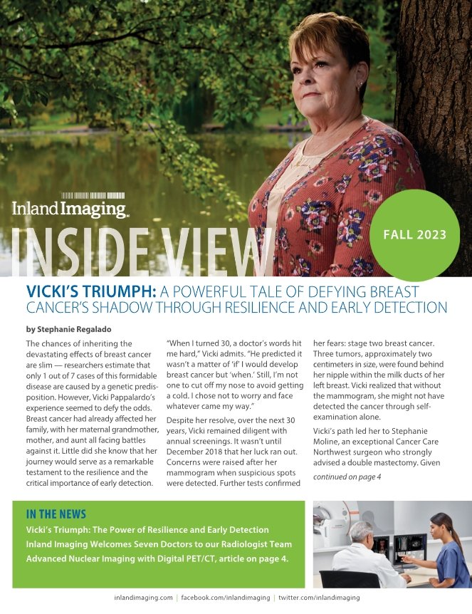
Vicki’s Triumph: The Power of Resilience and Early Detection
Inland Imaging Welcomes Seven Doctors to our Radiologist Team
Advanced Nuclear Imaging with Digital PET/CT
[SPOKANE, WA, October 5, 2023] Beginning on January 1st, 2024, patients who need medically necessary breast imaging will no longer have to pay out-of-pocket for those exams. The new law, recently passed by the Washington State Legislature eliminates cost-sharing for patients, including deductibles, coinsurance, and copayments for those services.
Exams covered under the new law include:
Follow-up breast imaging after an abnormal screening exam.
Imaging needed because a patient has detected a breast lump, has breast pain, or other symptoms.
Breast ultrasound or MRI exams when a patient is at high-risk for breast cancer.
"Currently, screening mammograms are cost-free to patients under the Affordable Care Act, because they’re considered preventive exams. For patients who need additional follow-up when something suspicious or abnormal has been detected we recommend additional follow-up exams, referred to as diagnostic imaging," said Dr. Amy Henkel, a breast imaging specialist with Inland Imaging.
“Under the current system, additional diagnostic imaging can be costly, causing many patients to put them off to avoid those expenses, delaying potential diagnosis and treatment of breast cancer. For many women, that means finding the cancer later, when it may be more serious and much harder to treat. We say, ‘early detection saves lives’ because it really does and the data proves it,” said Inland Imaging’s Dr. Henkel. “The new law makes it easier for patients to get follow-up coverage and ultimately, we believe it will save more lives.
NOTE: These changes in the law apply to patients covered by commercial health insurance plans. These benefits and changes in coverage may not apply to some of those covered by self-insured plans through their employers.
When you have questions about your health plan benefits and coverage, consult with your health insurance provider or the benefits administrator at your place of employment.

Dr. Allison Tillack Becomes a Certified B Reader
CBO Announces Leadership Transition, Introduces New CEO
Inland Imaging Announces Leadership Transition in the Tri-Cities
— August 2023
Inland Imaging is pleased to announce the promotion of John Crowley as Director of Operations at the company’s Tri-Cities’ imaging center in Kennewick. John replaces LuWanda (Louie) Leskinen, who is taking an extended leave to spend more time with her children and family. Leskinen previously served in technologist and team leader roles in both the CT and MRI departments before being named Director of Operations in the Tri-Cities, in 2021.
John Crowley began his medical imaging career in 1985, the year Inland Imaging opened its first outpatient facility near Sacred Heart Medical Center. He began as a Patient Care Assistant in the MRI department. John became a registered technologist specializing in Ultrasound in 1989 and added vascular Ultrasound in 1993.
John advanced to team leader in the Ultrasound Department after returning to inland following a year’s absence working for Advanced Technology Laboratories as an applications specialist in 1995. John was named Director of Ultrasound services in 2006. Crowley holds a master’s degree in healthcare management from SNHU.
— July 2023
Pat Atwal recently joined CBO in the role of Chief Executive Officer. Her priorities are to maintain the company’s established operational and service excellence and to position CBO for growth.
Pat is a local Spokane executive and entrepreneur who recently worked with INTRINIUM, a managed IT services provider. Her background is in technology, law, and leadership development. She is an attorney who transitioned to business operations roles and has been a COO, VP of Sales & Marketing, Director of Strategic Alliances, and other leadership roles. As COO of INTRINIUM she worked with large hospitals and local clinics to meet their information technology needs.
Kevin Kirk recently retired after 25 years of service with INLAND IMAGING and as Chief Operating Officer of CBO. He is looking forward to focusing on family life and devoting himself to spending time pursuing his passion for painting and fine art.
Jeneisa Sudbrink has been promoted to Chief Operating Officer. Over the past 7 years, her accomplishments at CBO have resulted in her taking on more responsibilities, moving from Manager to Director of Hospital Billing Operations, and to now COO.
ABOUT CBO
CBO is dedicated to solving the revenue cycle challenges of Critical Access Hospitals, Rural Healthcare Clinics, and individual rural practices. CBO’s experienced teams of billers, cash posters, customer service, and provider enrollment specialists improve the financial health of their clients by significantly reducing how long their accounts receivable are outstanding.
Find out more at getcbo.com, follow on LinkedIn (CBO, Central Billing Office Solutions for Healthcare), or reach out to Pat at patwal@inlandimaging.com.
— July 2023
Dr. Allison Tillack recently became a Certified B Reader. This unique designation requires specialized professional training and certification through the NATIONAL INSTITUTE FOR OCCUPATIONAL SAFETY AND HEALTH for demonstrating proficiency in reading diagnostic imaging associated with pneumoconiosis.
Pneumoconiosis is lung disease caused by the inhalation of airborne dust and fibers. Patients usually encounter these inhalants in the workplace environment, and therefore it is considered an occupational disease. The most common types of pneumoconiosis are asbestosis, silicosis, and coal miner’s lung. These particles cause inflammation and fibrosis in the lung resulting in irreversible lung disease. Prevention relies on workplace exposure management and health management regulations.
There are currently only 207 certified readers in the entire United States. Inland Imaging recently completed their first related imaging study at the HOLY FAMILY IMAGING CENTER with another one following closely behind it. This is the first time Inland Imaging has had a Certified B Reader on their professional radiology team and the first time the company has been able to offer this valuable service to occupational healthcare providers and their patients.
B Readers play an important role in providing medical diagnosis and follow up for workers with occupational exposures to airborne particulates such as coal mine dust, asbestos, and crystalline silica. Recently implemented Occupational Safety and Health Administration regulations have increased the need for the services provided by B Readers and INLAND IMAGING is proud to be able to offer those services due to Dr. Tillack’s special training and expertise.
Experts Agree Recommendations Don’t Go Far Enough. The USPSTF released new draft guidelines on Tuesday, May 9th. The task force says research indicates screenings beginning at age 40 could save 19% more lives.
However, many experts say the recommendations don’t go far enough. Organizations like the American College of Radiology, the American Society of Breast Surgeons, and the American College of Obstetricians and Gynecologists recommend women begin annual screenings at age 40 and that all women should have a risk assessment by age 25 to determine if they should begin screening earlier.
“We’ve long recommended annual screenings for breast cancer,” said Dr. Paige Flett, a breast imaging specialist at Inland Imaging. “It’s important for every woman, but especially for Black women, who are more likely to be diagnosed at an early age or with more aggressive cancers and are 40% more likely than white women to die of breast cancer.”
“Some women may benefit from additional breast cancer screening tests,” says Inland Imaging’s Dr. Amy Henkel. “Women at higher-than-average risk and those with dense breast tissue can consider supplemental screening by ultrasound or MRI in addition to annual screening mammography."
“Annual screening is important. The potential danger of missed cancers outweighs any inconveniences,” said Dr. John Bell, a breast imaging specialist at Inland Imaging. “Women diagnosed under age 50 are more likely to have aggressive cancers, and many who skipped mammograms during the pandemic and unfortunately are now seeing more serious diagnoses as a result. Annual screening means we can catch cancers sooner and save more lives.”
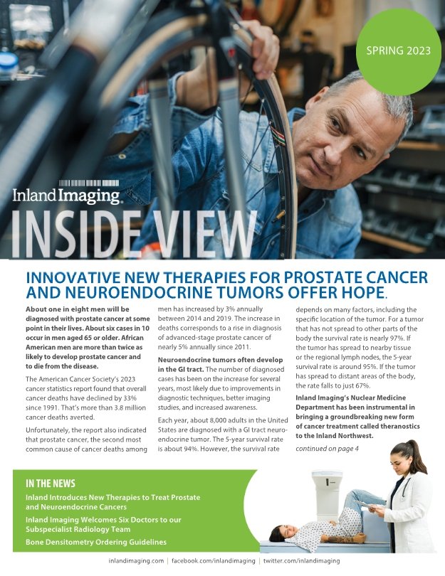
Inland Introduced New Therapies to Treat Prostate and Neuroendocrine Cancers
Inland Imaging Welcomes Six Doctors to our Subspecialist Radiology Team.
Bone Densitometry Ordering Guidelines
— April, 2023
About one in eight men will be diagnosed with prostate cancer at some point in their lives. About six cases in 10 occur in men aged 65 or older. African American men are more than twice as likely to develop prostate cancer and to die from the disease.
The American Cancer Society’s 2023 cancer statistics report found that overall cancer deaths have declined by 33% since 1991. That’s more than 3.8 million cancer deaths averted.
Unfortunately, the report also indicated that prostate cancer, the second most
common cause of cancer deaths among men has increased by 3% annually between 2014 and 2019. The increase in deaths corresponds to a rise in diagnosis of advanced-stage prostate cancer of nearly 5% annually since 2011.
Neuroendocrine tumors often develop in the GI tract. The number of diagnosed cases has been on the increase for several years, most likely due to improvements in diagnostic techniques, better imaging studies, and increased awareness.
Each year, about 8,000 adults in the United States are diagnosed with a GI tract neuroendocrine tumor. The 5-year survival rate is about 94%. However, the survival rate depends on many factors, including the specific location of the tumor. For a tumor that has not spread to other parts of the body the survival rate is nearly 97%. If the tumor has spread to nearby tissue or the regional lymph nodes, the 5-year survival rate is around 95%. If the tumor has spread to distant areas of the body, the rate falls to just 67%.
Inland Imaging’s Nuclear Medicine Department has been instrumental in bringing a groundbreaking new form of cancer treatment called theranostics to the Inland Northwest.
According to the Radiological Society of North America (RSNA), Theranostics refers to the pairing of diagnostic biomarkers with therapeutic agents that share a specific target in diseased cells or tissues.
More specifically, Theranostics describes the use of radioactive compounds to image specific disease targets and then to use that same tumor specific compound to deliver therapeutic radiation to that
specific targeted tumor type. Theranostics allows for a more targeted and personalized approach to cancer treatment by helping to provide customized targeted management for various cancers.
Inland Imaging currently provides two of these groundbreaking new forms of targeted cancer treatment: Pluvicto, first FDA-approved theranostic agent for treating metastatic prostate cancer;
and Lutathera, the first FDA-approved theranostic agent for treatment of advanced neuroendocrine tumors.
Currently, Inland Imaging is the only provider in the region offering both Pluvicto and Lutathera and one of only a handful of centers in the country able to offer advanced personalized dosimetric analysis to patients by employing SPECT/CT in combination with advanced software to verify the distribution of the therapy and quickly quantify tumor response during a patient’s treatment course. This allows Inland’s Theranostics team to tailor treatments to individual patients and subsequently reduces side effects by minimizing the radiation dose to normal tissue.
Inland Imaging is proud to provide these innovative therapies in Eastern Washington, Northern Oregon, Idaho, and Western Montana, which means more effective and advanced treatment, closer to home for those suffering from prostate or neuroendocrine cancers.
To learn more about Theranostics and the use of Lutathera and Pluvicto, talk to your doctor or go to Nuclear Oncology.
— March 2023
Bone Density (DXA) scans and Trabecular Bone Score (TBS) exams are important tools used to measure bone loss and the bone’s internal micro-architecture and strength. They are commonly used to diagnose osteoporosis and to assess an individual’s risk for osteoporotic fractures. The exam is simple, quick and noninvasive. It’s also the most commonly used and the standard method for diagnosing osteoporosis and bone fracture risk.
Patients must meet one or more of the following criteria for a screening DXA to be covered:
A woman at risk for osteoporosis who is estrogen deficient
A person whose X-rays show possible osteoporosis, osteopenia or vertebral fractures
A person taking prednisone or steroid-type medications, or is planning to take them
A person diagnosed with hyperpara thyroidism
A person being monitored during drug therapy
It is important that the criteria a patient meets be indicated on the DXA order.
Medicare and most insurance plans will only cover a Screening DXA once every two years. However, there are diagnoses that allow for the frequency of a DXA sooner than the 24 months. These include the following:
Cushing Syndrome
Osteoporosis without current pathological fracture (specifyanatomical site)
Long term (current) use of steroids
Long term (current) use ofbisphosphonates
Other specified disorders of bone density and structure (specify anatomical site)
Inland Imaging is pleased to announce the appointment of Jennifer Heimbigner as Chief Operations Officer (COO) of Inland Imaging Clinical Associates and Inland Imaging, LLC, the company’s outpatient imaging and clinical staffing divisions.
Heimbigner joins Inland Imaging after spending the last 23 years in increasingly responsible roles in healthcare administration with Cancer Care Northwest, most recently as CEO, a position she has held since 2017.
Jen started her career at Cancer Care Northwest as a front office receptionist and telephone operator. She was quickly promoted to Senior Administrative Assistant, and in 2010 became the clinic manager before moving to Director of Operations and then CEO. She obtained her undergraduate degree as well as her Master’s degree in Public Administration from Eastern Washington University.
“We are very excited to have Jen join our Inland family as she embodies our core values, strongly believes in caring for both her employees and patients, and will be an excellent cultural fit as well,” said Inland CEO Chris Patrick. “Jen has been described as a hands-on mentor who leads with compassion, ambition, and understanding.”
Jen has deep Spokane area roots, and enjoys family time at the lake and staying active through running and other outdoor activities. She is currently training to run her first full marathon in September.
INLAND IMAGING INTRODUCES BREAST IMAGING PATIENT NAVIGATOR. NEW ROLE TO HELP ASSURE CONTINUITY AND TRANSPARENCY IN PATIENT CARE — September 2022
Inland Imaging recently announced the appointment of Julie Smith to the role of Breast Imaging Patient Care Navigator.
The role of the breast imaging patient navigator is designed to provide Inland Imaging’s patients with a personalized path for their breast health. The Navigator supplies information and helps educate the patient on the various breast imaging modalities and services provided.
According to Smith, “I work with patients, referring clinicians, radiologists, medical specialists, community resources, and staff to help meet the needs of each patient within their breast imaging care journey, from screening mammography through diagnosis.”
Smith has been involved in breast imaging for over thirty years. She has been with Inland Imaging for twenty-five years and has served for over twenty years as a team leader in mammography. According to Julie, “I have a passion for breast imaging work and truly enjoy the opportunity to assist and make connections with my patients.”

INTRODUCING LUTATHERA: A REVOLUTIONARY NEW SERVICE LINE FOR THE TREATMENT OF CANCER. - Inland Imaging recently announced the introduction of LUTATHERA, a prescription treatment for adults with specific types of cancer.
INLAND IMAGING INTRODUCES BREAST IMAGING PATIENT NAVIGATOR. NEW ROLE TO HELP ASSURE CONTINUITY AND TRANSPARENCY IN PATIENT CARE.
Inland Imaging is pleased to announce the appointment of Jennifer Heimbigner as Chief Operations Officer (COO) of Inland Imaging Clinical Associates and Inland Imaging, LLC, the company’s outpatient imaging and clinical staffing divisions.
Inland Imaging Welcomes Six Doctors to our Subspecialist Radiology Team.

MY MAMMOGRAM EXPERIENCE - My doctor told me to get a screening mammogram
INLAND IMAGING INTRODUCES ILLUCCIX®: A Radioactive Diagnostic Agent for PET-CT
Inland Imaging Welcomes Doctors Cox, Lee, and Penna to Our Subspecialist Radiology Team
WHERE'S THE LEAD APRON? - Shielding is Now Discouraged During Your X-Ray
WHERE'S THE LEAD APRON?
Shielding is Now Discouraged During Your X-Ray - June 1, 2022
When you visit Inland Imaging for an X-ray exam, you may notice that we no longer ask patients to wear shielding. Based on over 70 years of research, we now know that keeping you healthy and safe doesn’t require the use of shields. With modern X-Ray technology, lead aprons do not benefit the person being imaged. They may obscure the area we are trying to see, and in some cases, interfere with X-ray equipment. This is true for individuals of all ages. Regulations still require staff and non-patients in the exam room to wear lead aprons.
The following organizations support this practice update:
American Association of Physicists in Medicine (AAPM)
American Board of Radiology (ABR)
American College of Radiology (ACR)
American Society of Radiologic Technologists (ASRT)
Image Gently & Image Wisely
Society for Pediatric Radiology (SPR)
Interested in Learning More?
Visit this website:
https://www.radiologyinfo.org/en/info/safety-patient-shielding
INLAND IMAGING Introduces Illuccix®: a Radioactive Diagnostic Agent for PET-CT (positron emission tomography)
Improves the Detection and Treatment of Prostate Cancer. — May 30, 2022
Recently approved for use by the FDA, the availability of this new agent helps improve access to more advanced and detailed imaging for patients facing diagnosis and treatment of prostate cancer.
“The approval of Illuccix will give patients considerably improved access to PSMA-PET imaging. PSMA PET can be used for initial staging of prostate cancer or evaluating recurrent disease,” said David Mariner, Director of Nuclear Medicine and PetCT at Inland Imaging. “PSMA-PET will also
be used in conjunction with a new prostate cancer therapy, Lu-177 PSMA, that we anticipate starting later this summer.”
Illuccix is the first commercially available FDA-approved product to enable wide accessibility to PSMA-PET imaging for physicians and eligible patients in the region.
“This product offers a level of sensitivity and specificity that is superior to regular FDG PET for most prostate cancer patients and may help us provide better patient experiences, and outcomes, as a result,” said Dr. Irene Cruite, a radiologist at Inland Imaging specializing in Nuclear Medicine.
“Improved imaging can provide physicians with the insights to more accurately determine the current state of disease and the most appropriate treatment
pathway. It gives patients access to a specific and sensitive imaging tool for the detection of prostate cancer throughout the body.”
According to the American Cancer Society, prostate cancer is the second leading cancer in men in the United States with nearly 250,000 cases and more than 34,500 deaths from the disease in 2021 — a significantly higher incidence than either lung cancer (119,000 new cases) or bowel cancer (80,000 new cases). More than 800,000 U.S. men are estimated to be living with prostate cancer today.
CEO Steve Duvoisin Announces Leadership Transition
ABUS Improves Early Cancer Detection for Dense Breasts
Inland Imaging Welcomes Five Radiologists to our Spokane and Tri-Cities’ Teams
CT Coronary Artery Screening Exam offered for $150
Inland Contrast Reaction Training

Spokane, WA – INLAND IMAGING’s long-time CEO, Steve Duvoisin recently announced his retirement along with upcoming changes to the company’s leadership team.
Effective April 1, 2022, Chris Patrick, currently CEO of Inland Imaging subsidiary Nuvodia, a national professional services company, will assume the role of CEO for Inland Imaging’s business organizations (Inland Imaging, LLC - Outpatient Imaging joint venture with Providence, Inland Imaging Business Associates – Business Services, Inland Imaging Clinical Associates – Staff Leasing Services and Inland Imaging Investments - strategic investment company).
Also effective April 1, 2022, Sarah Russell, currently COO of Inland Imaging PS, the company’s professional radiology group, will assume responsibilities as CEO of that organization whose 120+ radiologists practice in Seattle, Spokane, Tri-Cities, Walla Walla and Missoula, as well as many other locations throughout Washington, Montana, Idaho and Utah.
Russell joined Inland Imaging in 1997 and is responsible for all aspects of the professional radiology practice. According to Duvoisin, “Sarah has been instrumental in the growth of the practice and the successful expansion of its operations. She has overseen the group’s growth from 14 to more than 120 radiologists, and has been an integral part of creating a great culture and helping to chart its future course. We look forward to her continued success in her new role as the CEO of Inland Imaging, PS.”
Patrick’s background includes more than 25 years of diverse professional experience, including positions as market director at AT&T, COO of Quick Study Radiology, as well as a number of high level positions in the information technology sector. “As CEO of Nuvodia, Chris has guided the company’s growth and award winning culture, working across multiple industries, including healthcare, professional services and energy,” said Duvoisin. “We are looking forward to seeing Chris apply his leadership and competitive spirit to growing Inland‘s various organizations and addressing the challenges our organizations will face as we look to the future.”
Duvoisin has been CEO of Inland Imaging’s professional radiology group and outpatient imaging company, as well as its business services, information technology, strategic planning and investment organizations for the last 37 years. Steve joined the company in 1984 when it opened its first outpatient-imaging center with six radiologists and 30 clinical and clerical employees. He serves on a variety of professional, civic, philanthropic, and arts-related boards and advisory groups. Steve is looking forward to spending more time with his family in the future.

Inland IR Specialists Join National Research Study
Dayton General Hospital Selects Inland Imaging
Providence St. Mary’s Radiologists Join Inland Imaging
Inland Imaging Welcomes Five Radiologists to our Team
Inland Sponsors the 5th Annual
Every Woman Can Pink Ribbon Run
October 3
By Natasha Nellis at the Spokane Journal of Business
Inland Imaging LLC has enrolled the first patient in a new liver cancer treatment trial.
Using what’s known as Y-90 radiation therapy, the treatment uses microscopic beads filled with yttrium-90 isotope “designed to be delivered directly into tumors through the arteries that feed the tumors,” says Dr. Douglas Murrey, an interventional radiologist at Inland Imaging. Once in place, the radioactive isotope destroys the surrounding tissue from within the tumor.
Read the rest of the article at the Spokane Journal of Business.

Ultrasound and MR Liver Elastography: Minimally Invasive, Fast, and Convenient Liver Elastography Offers Patients a Pain-Free Alternative to Invasive Liver Biopsies
Inland Welcomes Dr. Dunn to Our Radiologist Team
COVID-19 Update
Choosing the Right Scanner for Your MRI Exam
March 2021 —
Often, people with liver fibrosis don’t experience any signs or symptoms. Chronic liver disease, most commonly Hepatitis C, Hepatitis B, alcohol abuse, and non-alcoholic steatohepatitis, eventually results in liver scarring. When this scarring, or fibrosis, becomes severe, the risk of complications of chronic liver disease, including liver cancer, increases dramatically. Inland Imaging’s Ultrasound (US) and Magnetic Resonance Elastography (MRE) studies can help provide the answers you and your physician need to guide your care.
Effective and Painless Alternative to Invasive Liver Biopsies
Liver biopsy is useful in determining the severity of liver fibrosis. However, an invasive biopsy also invites potential medical complications as well as a variety of other inconveniences including pain, the need for sedation, arranging for transportation, and as much as a full day of recovery time.
Liver elastography can provide all the information providers need without the inconvenience and expense of a biopsy in most patients. The patient only needs to undergo a non-invasive ultrasound or MRI exam.
What to Expect During the Exam
ULTRASOUND LIVER ELASTOGRAPHY: The exam itself is simple and painless.
To prepare, patients should not eat or drink for 4 hours prior to their appointment. During the exam, the patient lies comfortably on their back while an ultrasound technologist takes images of the liver, gallbladder, spleen, and sometimes more, depending on the ordering provider’s request. An additional five to ten minutes of imaging the liver helps obtain the required elastography measurements. After the exam, the patient can leave with no lingering side effects.
MAGNETIC RESONANCE ELASTOGRAPHY (MRE): The usefulness of ultrasound (US) elastography is limited in larger patients, who have a higher degree of success with MRI vs. US. MRE is done with the same type of scanner and many of the same steps as a traditional MRI exam.
During an MRE procedure:
The patient reclines, face-up on the examination table.
A radiology technologist will place a small pad on the patient’s abdomen. The pad emits vibrations that pass through the liver.
The table slides into the MRI scanner. Patients may be given earplugs or headphones before the test to help block the noise of the scanner, which can be loud.
Once inside the scanner, the pad activates and measurements are recorded onto a computer and turned into a visual map of the liver.
The scan takes under 30 minutes to complete.
Findings and Follow-up
A radiologist reviews the images and assesses an “F-score,” or fibrosis score describing the level of liver scarring present. The Radiologist then generates a report containing the F-score information typically within 24 hours after completion of the exam. Your physician can then use this information to help determine the most appropriate course of care — all while more effectively
controlling health care costs.
To find out if you’re a candidate for Liver Fibrosis assessment using ultrasound or MR Elastography, ask your primary care provider or contact inland imaging.
In Spokane call (509) 455.4455. In Tri-Cities call (509) 374.4030.
Magnetic Resonance Imaging (MRI) is one of the safest, most comfortable imaging techniques available. The sophisticated technology combines the use of a powerful magnetic field, radio waves, and a specialized computer system to produce detailed, multi-dimensional images of inside your body.
One advantage of an MRI scan is its ability to produce exceptionally detailed images of almost any part of the body, including internal organs, muscles, nerves, blood vessels and small soft tissues around the joints or spine that cannot always be seen easily using other imaging methods. Choosing Inland Imaging for your MRI exam offers you choices to help make your scan as comfortable and effective as it can be.
OPEN MRI
Inland’s Open MRI Scanner, is specially designed for those who experience claustrophobia or anxiety in small spaces. This machine features a spacious opening with a 270-degree view. Patients are even able to maintain physical contact with a friend or family member during the exam if they choose. The platform can be lowered
to wheelchair height, offering easier access for those with limited mobility. This is an excellent option for those who have concerns about initial comfort when scheduling an MRI exam.
EXTREMITY MRI
In addition to the Open MRI, patients who don’t require a whole-body exam can take advantage of an Extremity MRI exam option. The extremity MRI is a specialty scanner for patients needing an exam of the arm (including elbow, wrist, and hand) or the leg (including knee, ankle, and foot). This exam option provides both outstanding image quality and a more relaxed and enjoyable experience for the patient.
3T (TESLA) MRI
The strength of the magnetic field generated by an MRI is measured in units known as Teslas. Named after the brilliant inventor, these units of measure define the quality of the images an MRI can produce. Simply put, the more Teslas, the higher quality the images. The technology of the 3T MRI scanner addresses everything from routine medical exams to clinical research. This model is top of its class.
To find out which of our amazing MRI scanners is the best choice for your exam, ask your physician, or call Inland Imaging at (509) 455.4455.
September 30, 2020 —
The Interventional Radiology team moved on September 1, 2020, to a new space adjacent to our recently opened Outpatient Based Interventional Center (OBIC) on the campus of Providence Holy Family Hospital in North Spokane.
This new location offers our highly trained and experienced team the opportunity to serve our providers and their patients with the highest levels of treatment and care available in our region.
We offer an interdisciplinary approach collaborating with local and regional specialists to provide state-of-the-art care, inpatient and outpatient consults, procedures, and follow-up care for a wide variety of medical conditions.
We look forward to serving you from our new Northside location and welcome your feedback on how we can make your experience with our practice the best it can be.
October 20, 2020 —
Each year, breast cancer detection technology delivers more detailed and accurate results. Nonetheless, more than 40,000 U.S. women still die from breast cancer each year.
Nearly half of all women in their 40s and 50s have dense breasts—which can be more difficult to read using standard mammography. Today, there’s more and more evidence that women with dense breasts who are at average risk for breast cancer, could benefit from MRI screening as well.
Abbreviated MRI (AB-MRI), takes about 10 minutes and delivers results that other, less sensitive exams, might miss. AB-MRI has been demonstrated in clinical studies to detect up to 16 per 1,000 more invasive cancers than mammography alone.
A recent study reported in the Journal of the American Medical Association compared AB-MRI with standard digital 3D mammography. It found that AB-MRI detected significantly more cancers in women with dense breasts. The study’s most important conclusion may be that AB-MRI appears to catch more aggressive cancers earlier, when they are most treatable.
Is Abbreviated MRI Right for You?
Inland Imaging is now offering women the option of AB-MRI for $500. The exam is not currently covered by insurance. Talk with your OBGYN or primary care provider to explore your options and find out if AB-MRI might be right for you.
To learn more, go to https://www.inlandimaging.com/breast-imaging-subspecialties and mammographysaveslives.org.
[Spokane, WA] SEPTEMBER 15, 2020. Recently, Inland Imaging acquired and installed a new, next-generation digital PET/CT scanner — the Biograph Vision, designed and manufactured by Siemens Healthineers. The new scanner — the first Siemens digital PET/CT to be installed on the West Coast — leverages advanced digital technology to help Inland Imaging’s sub-specialized radiologists detect small, disease related lesions earlier, better quantify them, and more fully understand disease progression and response to treatment.
“Inland Imaging’s new digital PET/CT scanner represents the next-generation of technology. It is the most advanced and most sensitive PET/CT technology available in our region today, designed to deliver an exciting new level of precision molecular imaging, and improved patient experience,” said Dr. Irene Cruite, the Nuclear Medicine Section Head at Inland Imaging. “This new scanner takes a technological leap forward in quality and efficiency. It has improved our ability to deliver the answers patients and their health care providers need to provide accurate and timely diagnosis, stage disease, determine the best course of treatment, and monitor disease progression and response to treatment. The digital capability enhances our ability to detect and characterize small lesions, and it increases conspicuity of subtle areas of disease that would otherwise not be apparent. It is also synergistic with new radionuclide therapies, new PET/CT imaging radiotracers, and new applications of current radiotracers. In addition, the new scanner is designed with a series of features that allow for metal artifact reduction, lower patient radiation exposure and shorter scan times for patient comfort without impacting image quality. Its wide-bore design also helps improve patient experience and comfort. This new technology has enhanced our ability to deliver value-oriented care and potentially improve patient outcomes by getting the right treatment to the right patient at the right time.”
THE DIGITAL DIFFERENCE
“Digital PET/CT clearly surpasses analog scanners in terms of image quality, detail, and sharpness,” said David Mariner, manager of Inland Imaging’s Nuclear Medicine team. “With better spatial resolution and increased sensitivity, this technology delivers much higher image quality using less radiation and shorter scan times when compared to older analog scanners.
More detailed image quality translates into more accurate diagnoses, allowing patients and their providers to take advantage of the most effective early treatment options available. We are scanning using 40-70% less radioactive isotope than would be used with an older analog scanner, which significantly reduces patient radiation exposure. In some cases we have successfully scanned patients at half the radiation dose that similar groups around the country consider to be their low dose protocol on newer scanners. We have also reduced scan time by 20-40%, which minimizes patient motion during image acquisition and improves patient comfort. The new scanner also allows us harness new PET/CT applications and new radionuclide therapies, thereby providing leading-edge services to our patients and their health care providers.”
“We are excited to be the first to bring this new digital PET/CT technology to the region,” said Bob White, chief operating officer at Inland Imaging. “Its high resolution and enhanced imaging quality allow our highly trained radiologists to identify small lesions much earlier. By incorporating the capabilities of this advanced digital PET/CT scanner into our practice, we can improve patient safety and comfort, identify disease sooner, improve outcomes, reduce costs, and help patients take advantage of the most effective therapies.”

OBIC Achieving Excellent Outcomes
Inland Imaging Welcomes Three Radiologists to Our Team —Jordan Castle, MD, Wendy Ehieli, MD and Richard Kennard, MD
COVID-19 Update
Centers Earn Vascular Accreditation by the IAC
Inland Imaging’s Holy Family, South Cowley, and Spokane Valley Centers Earn Vascular Testing Accreditation by the IAC
Early detection of life-threatening heart disorders, stroke and other diseases is possible through the use of vascular testing procedures performed within hospitals, outpatient centers and physicians' offices. Cardiovascular diseases are the No. 1 cause of death in the United States. On average, one American dies every 39 seconds of cardiovascular disease - disorders of the heart and blood vessels. Stroke, a disorder of the blood supply to the brain, is the third leading cause of death and the leading cause of disability in the country, with nearly 800,000 new strokes occurring annually.
There are many factors that contribute to an accurate diagnosis based on vascular testing. The training and experience of the technologist performing the procedure, the type of equipment used and the quality assessment metrics each facility is required to measure, all contribute to a positive patient outcome. IAC accreditation is a "seal of approval" that patients can rely on as an indicator of consistent quality care and a dedication to continuous improvement.
Inland Imaging’s Holy Family, South Cowley, and Spokane Valley Centers have been granted a three-year term of accreditation by the Intersocietal Accreditation Commission (IAC) in Vascular Testing in the area(s) of Extracranial Cerebrovascular Testing, Peripheral Venous Testing, Peripheral Arterial Testing.
Accreditation by IAC indicates that Inland Imaging has undergone an intensive application and review process and is found to be in compliance with the published Standards, thus demonstrating a commitment to quality patient care in vascular testing. Comprised of a detailed self-evaluation followed by a thorough review by a panel of medical experts, the IAC accreditation process enables both the critical operational and technical components of the applicant facility to be assessed, including representative case studies and their corresponding final reports.
Decades-long Drop in Breast Cancer Death Rate Continues
October 30, 2019 —
Mortality decline has slowed in recent years. Breast cancer now leading cause of cancer death for black women in six states.
A decades-long decline in the breast cancer death rate continues, but has begun to slow in recent years, while breast cancer incidence rates continue to inch up.
Breast cancer is the most common cancer (excluding skin cancers) diagnosed among U.S. women and is the second leading cause of cancer death among women after lung cancer. In 2019, approximately 268,600 new cases of invasive breast cancer will be diagnosed among U.S. women, and 41,760 women will die from the disease.
The overall breast cancer death rate has decreased consistently since 1989,
attributed both to improvements in early detection (through screening as well as increased awareness of symptoms) and treatment for a total decline of 40 percent through 2017. As a result of this decline, 375,900 breast cancer deaths have been averted in U.S. women through 2017.
The latest data shows that the pace of the mortality decline has slowed in recent years — from a drop of 1.9 percent per year during 1998 through 2011 to 1.3 percent per year during 2011 through 2017, largely driven by the trend in white women. Consequently, the black-white disparity in breast cancer mortality that widened over the past three decades has remained stable since 2011. Nevertheless, in the most recent period (2013-2017), the breast cancer death rate was 40 percent higher in black women versus white women, despite slightly lower incidence rates. This disparity is magnified among black women under 50, among whom the death is rate double that of white women.
In the most recent 5-year period (2013-2017), the breast cancer death rate declined by 2.1 percent per year in Hispanics/Latinas, 1.5 percent per year in blacks, 1 percent per year in whites, and 0.8 percent per year in Asians/Pacific Islanders, and was stable in American Indians/Alaska Natives. However, mortality rates are no longer declining for black women in Colorado and Wisconsin and for white women in Nebraska, Texas, and Virginia.
To find out more about breast cancer detection, go to inlandimaging.com/breast-imaging, or call to schedule your annual screening mammogram at (509) 455-4455.
Early detection saves lives.
Inland Imaging Earns IAC Vein Center Accreditaion
November 2019 —
Inland Imaging recently completed the accreditation process through the Intersocietal Accreditation Commission for our vascular labs at Providence Holy Family Hospital, and our South Cowley, and Valley Imaging Centers.
The Intersocietal Accreditation Commission (IAC) accredits vein centers that perform evaluation and management of superficial venous disorders. IAC accreditation is a means by which facilities can evaluate and demonstrate the level of patient care they provide. IAC Vein Center accreditation is granted for a three-year period from the original date of decision.
MOST CURRENT AND FORMER HEAVY SMOKERS AREN’T BEING SCREENED FOR LUNG CANCER.
INLAND EXPANDS IN NORTH SPOKANE
LIVER ELASTOGRAPHY: AN ALTERNATIVE TO INVASIVE BIOPSY
VIRTUAL COLONOSCOPY: A LESS INVASIVE WAY TO SCREEN FOR COLON CANCER
Advanced MRI Technology can now show detailed images of the heart.
APRIL 2019 — Magnetic Resonance Imaging (MRI) uses magnetic fields and radio waves to generate images of the body. MRI has become a very valuable diagnostic tool detecting everything from cancer, heart and vascular disease, strokes, and disorders of the joints and musculoskeletal system. MRI often also allows physicians to avoid unnecessary surgery and more invasive diagnostic procedures. MRI technology produces extremely
detailed images of body tissue, organs and bones without the need for radiation.
A Cardiac MRI exam at Inland Imaging offers a clear window into the heart and vascular system, allowing physicians to view—with exceptional detail—the function and structure of the heart, heart chambers, valves and major vessels.
This information helps physicians diagnose and treat a variety of cardiovascular conditions. For example, a Cardiac MRI may help detect and evaluate coronary artery disease or
defects with the heart chambers or valves. It is also used to determine the extent of damage caused by a heart attack or progressive heart disease and to measure the buildup of plaque and blockages in the blood vessels.
If you’re concerned about heart health, talk with your physician to see if the answers a Cardiac MRI will provide might be of value in determining the steps you can take to improve your overall cardiac fitness and health.